- 15″ color CRT monitor
- Pulse Wave Doppler
- Color Doppler Flow Imaging
- Power Doppler Flow Imaging
- Directional Power Doppler Flow Imaging
- Tissue Harmonic Imaging
- Built-in imaging archive
- CD-R/W and USB ports
- Measurement & calculation software packages

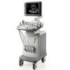
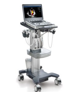
Mindray DC-6 Ultrasound Machine
National Ultrasound, the North American Distributer for Mindray, stands behind the Mindray DC-6 Ultrasound Machine, a color Doppler system for general imaging applications. The DC-6 incorporates the latest in digital ultrasound image processing technologies, providing advanced clinical solutions. This machine creates precise 2D color Doppler images and features fine tissue optimization to eliminate noise. Features include multi-beam parallel imaging, vessel imaging, and more. The DC-6 creates an interactive process connecting sonographers, patients, and the system. Its ergonomic design and powerful hardware greatly increase diagnostic accuracy and efficiency. The DC-6 is no longer manufactured, IDD recommends the DC-N3 or DC-70 to upgrade too. Contact us today to find which system meets your needs.
Get your questions answered about the Mindray DC-6 Ultrasound Machine by our trained National Ultrasound staff. Fill out the quick quote form below to start the process.
- 3C5A – Convex array transducer 3C5A (2.5/3.5/5.0/H4.6/H6.0MHz)
- 7L4A – Linear array transducer (5.0/7.5/10.0MHz)
- 6CV1 – Micro-convex endocavity transducer (5.0/6.5/8.0MHz)
- 3C1 – Micro-convex array transducer (2.5/3.5/5.0/H4.6/H6.0MHz)
- 7L6 – Linear array transducer (5.0/7.5/10.0MHz)
- 7L5 – Linear array transducer (5.0/7.5/10.0MHz)
- 10L4 – Linear array transducer (8.0/10.0/12.0MHz)
- 6C2 – Micro-convex array transducer (5.0/6.5/8.0MHz)
- 2P2 – Phased array transducer (2.0/2.5/3.0/H3.5/H4.0MHz)
- 6LE7 – Intrarectal linear array transducer (5.0/6.5/8.0MHz)
- 7LT4 – Intraoperative T-type linear array transducer (5.0/7.5/10.0MHz)
- 6LB7 – Intrarectal biplanar transducer (5.0/6.5/8.0MHz)
- DICOM 3.0
- Water resistant footswitch
- ECG module
- Needle guides
- Printer
Related Products
Ultrasound Machines

Ultrasound Machines
Ultrasound Machines
Ultrasound Machines